
Achalasia of the Esophagus
Achalasia is a term of Greek origin meaning “failure of relaxation.”
Achalasia is a rare primary esophageal motility disorder characterized by the absence of peristalsis and impaired relaxation of the lower esophageal sphincter (LES), resulting in impaired food transit and esophageal stasis.
In 1672, the English physician Sir Thomas Willis described cardiospasm and its treatment using dilation with a sponge attached to a whalebone. Two hundred forty-one years later, on April 14, 1913, the German surgeon Ernest Heller performed the first successful esophagomyotomy. In 1937, F. C. Lendrum proposed that functional esophageal obstruction was caused by failure of LES relaxation, after which the term “cardiospasm” was replaced by “achalasia.” In 1962, Dor reported performing an anterior partial fundoplication using his own technique, and in 1963 André Toupet described posterior partial fundoplication. In 1991, Shimi and colleagues in the United Kingdom performed the first laparoscopic Heller myotomy, 77 years after the original procedure.
The epidemiology of achalasia is not fully understood. The global incidence is estimated at 0.5–1.0 cases per 100,000 population per year. The disease can occur at any age, with a peak incidence between 30 and 60 years, and is extremely rare during the first two years of life. For example, at the University of California, San Francisco (UCSF), the mean age at diagnosis was 48 years. Achalasia affects men and women equally, with no ethnic predilection. Familial cases have been reported but account for less than 1% of all cases described in the literature. The triple-A syndrome (achalasia–alacrima–ACTH-resistant adrenal insufficiency), also known as Allgrove syndrome, is a rare condition. Although achalasia is associated with an increased risk of esophageal cancer, long-term studies indicate that it does not significantly affect life expectancy. In one study, the mean age at death among patients with achalasia was 80 years, suggesting a relatively low incidence of esophageal cancer.
PATHOGENESIS AND ETIOLOGY
Motor innervation of the esophagus is mediated by the vagus nerve through the submucosal (Meissner’s) plexus (Fig. 1A). Esophageal innervation differs between the proximal and distal segments.
The striated muscles of the proximal esophagus are innervated by somatic efferent fibers of the vagus nerve (Fig. 1A). In contrast, the smooth muscle of the distal esophagus is innervated by preganglionic vagal fibers originating from neuronal cell bodies located in the dorsal motor nucleus.
Preganglionic fibers primarily innervate the submucosal plexus via cholinergic pathways. The esophageal wall and the lower esophageal sphincter (LES) are further innervated by postganglionic neurons, which consist of both excitatory and inhibitory neurons.
Postganglionic excitatory neurons release acetylcholine, whereas inhibitory neurons release nitric oxide (NO) and vasoactive intestinal peptide (VIP), which mediate contraction and relaxation of the esophagus and the lower esophageal sphincter, respectively (Fig. 1B).
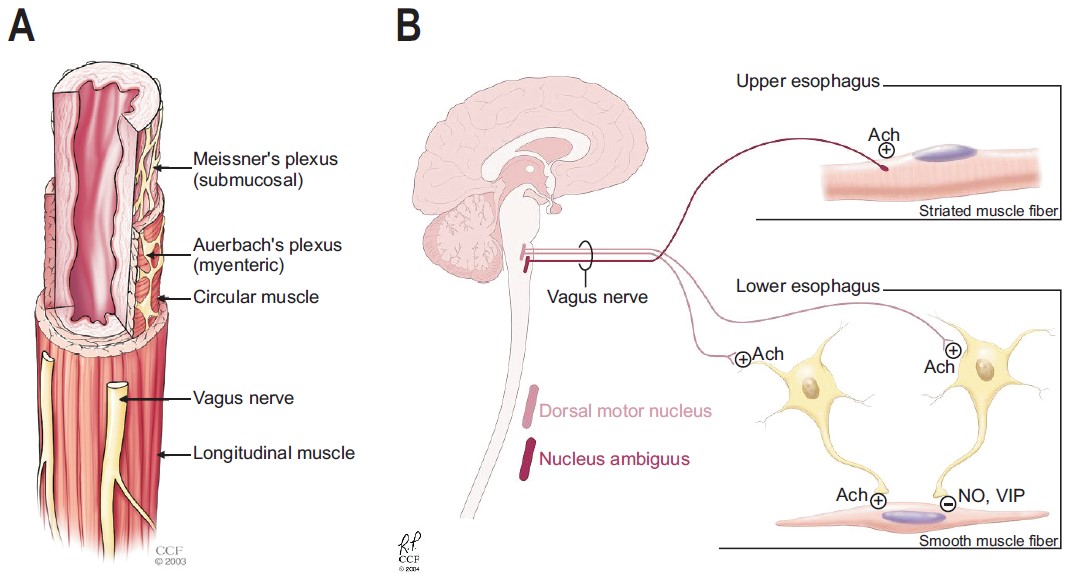
Figure 1.
Source: Ates F., Vaezi M.F. The Pathogenesis and Management of Achalasia: Current Status and Future Directions. Gut and Liver. 2015;9(4):449–463. https://doi.org/10.5009/gnl14446.
In addition to tonic contraction and relaxation, inhibitory neurons play a crucial role in maintaining normal esophageal peristalsis. At rest, the esophagus remains in a contracted state; however, during swallowing, inhibitory neurons are activated to suppress excitatory neuronal activity, resulting in esophageal relaxation.
Peristalsis represents the net effect of coordinated relaxation and contraction mediated by inhibitory and excitatory neurons of the submucosal plexus along the entire length of the esophagus.
In achalasia, there is a loss of nitric oxide (NO) and vasoactive intestinal peptide (VIP) released from inhibitory neurons. Consequently, the loss of inhibitory innervation leads to impaired relaxation of the lower esophageal sphincter (LES) as well as complete absence of esophageal peristalsis.
From a pathophysiological perspective, the loss of inhibitory innervation may result from both central and peripheral mechanisms. Central causes include lesions of the central nervous system, particularly involving the dorsal motor nucleus of the vagus nerve. Peripheral mechanisms involve the loss of inhibitory ganglion cells within the esophageal enteric (submucosal) plexus.
Kimura was the first to propose that central nervous system lesions could account for the clinical and manometric abnormalities observed in achalasia. In 1929, he examined histological sections from postmortem specimens of three patients with achalasia and identified degeneration of neuronal cells within the dorsal motor nucleus of the vagus nerve.
Similar findings were later reported by Cassella. His group performed histological analysis of serial brainstem sections from two patients with achalasia and one control subject. They demonstrated a bilateral reduction in neuronal count within the dorsal motor nucleus of the vagus nerve ranging from 34% to 43% compared to controls.
To validate these observations, Higgs experimentally induced bilateral lesions of the dorsal motor nuclei of the vagus nerve in 13 cats using direct electrical current. Nine of the 13 animals (69%) developed manometric and radiological features consistent with achalasia.
These findings suggest that lesions within the central nervous system may contribute to the development of manometric manifestations characteristic of achalasia.
Abnormalities in vagal nerve fibers outside the central nervous system have also been associated with achalasia. Using electron microscopy, Cassella identified vagal nerve abnormalities resembling Wallerian degeneration in patients with achalasia. Additionally, in a patient who underwent highly selective vagotomy for recurrent bleeding from a duodenal ulcer, esophageal manometry revealed findings consistent with achalasia. However, since most patients undergoing vagotomy do not develop symptoms of achalasia, this observation is likely an isolated atypical case.
It is also possible that the observed alterations in vagal nerve fibers and degeneration of neurons in the dorsal motor nucleus are secondary phenomena resulting from loss of trophic interaction with the target organ, namely the esophageal enteric (submucosal) plexus. Overall, extrinsic innervation abnormalities are rare in achalasia and are unlikely to represent the primary pathogenic mechanism.
Current evidence suggests that the principal neuronal defect in achalasia is an imbalance between excitatory and inhibitory neurons within the enteric nervous system. Preservation of cholinergic excitatory neurons was demonstrated by Holloway in a case- control study involving 27 patients with achalasia and 21 healthy controls. Both cholinergic and anticholinergic agents were administered, followed by esophageal manometry.
Anticholinergic agents reduced lower esophageal sphincter pressure in both groups, whereas cholinergic agents increased it, confirming the preservation of cholinergic excitatory pathways in achalasia. This mechanism also underlies the therapeutic effect of botulinum toxin, which reduces LES pressure by inhibiting acetylcholine release.
In contrast to preserved excitatory innervation, numerous physiological studies demonstrate absent or impaired inhibitory innervation in achalasia.
Dodds conducted a case-control study in which 24 patients with achalasia received a bolus dose of cholecystokinin octapeptide. The control group, consisting of seven healthy volunteers and 32 patients without idiopathic achalasia undergoing manometry, received the same intervention.
In the control group, stimulation of both inhibitory neurons and smooth muscle of the LES resulted in a net effect of sphincter relaxation, as inhibitory neuronal activity predominates over direct smooth muscle stimulation.
In contrast, in patients with achalasia, administration of cholecystokinin octapeptide produced a paradoxical increase in LES pressure. This is attributed to the absence of inhibitory neurons, allowing unopposed direct stimulatory effects on smooth muscle (Fig. 2), thereby highlighting the underlying inhibitory deficit.
Therefore, this test may be clinically useful in patients with post-fundoplication dysphagia and suspected achalasia. An increase in basal LES pressure following administration of cholecystokinin octapeptide strongly supports the diagnosis of achalasia.
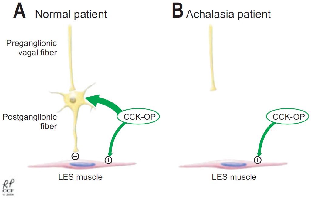
Figure 2. Source: Ates F., Vaezi M.F. The Pathogenesis and Management of Achalasia: Current
Status and Future Directions. Gut and Liver.. 2015;9(4):449–463. https://doi.org/10.5009/gnl14446
The loss of inhibitory neurons as a primary pathological mechanism in idiopathic achalasia is further supported by studies investigating inhibitory neurotransmitters. Vasoactive intestinal peptide (VIP), an inhibitory neurotransmitter of the esophageal submucosal plexus, has been shown to induce smooth muscle relaxation in vitro and promote relaxation of the lower esophageal sphincter in vivo.
Subsequent studies demonstrated that nerve fibers normally present in the submucosal plexus and the lower esophageal sphincter are either absent or significantly reduced in patients with achalasia.
More recent evidence identifies nitric oxide (NO) as the principal inhibitory neurotransmitter within the esophageal enteric nervous system. Animal studies have demonstrated that NO plays a critical role in regulating esophageal neuromuscular function, including LES relaxation and normal peristalsis.
A significant reduction in NO-containing neurons has been observed in patients with achalasia. Furthermore, pharmacological inactivation of NO in healthy volunteers using recombinant human hemoglobin resulted in manometric findings similar to those observed in achalasia.
NO-producing neurons and VIP-containing neurons typically coexist within the esophageal submucosal plexus, and their simultaneous loss likely underlies the clinical manifestations of achalasia.
The presence of familial cases suggests a potential genetic component in the pathogenesis of achalasia. Familial clustering is most commonly reported in pediatric populations, particularly among siblings and, in some cases, monozygotic twins. Parent–child associations have also been described.
Although these observations suggest a possible autosomal recessive inheritance pattern, the rarity of familial cases does not support a major etiological role for genetic factors in most patients. Instead, genetic predisposition may increase susceptibility to environmental triggers that contribute to disease development.
Several studies have proposed a potential association between viral infections and achalasia. Elevated serum antibody levels against measles and varicella-zoster viruses have been reported in patients with achalasia compared to controls. However, not all individuals exposed to these viruses develop achalasia.
Polymerase chain reaction studies have failed to detect viral genetic material in esophageal tissue. Even in studies reporting viral presence, a causal relationship has not been established. Overall, current evidence does not support an infectious etiology for achalasia.
An exception is Chagas disease, caused by Trypanosoma cruzi, which closely mimics the pathophysiology of primary achalasia.
An increased prevalence of circulating antibodies against the esophageal myenteric plexus has led to the hypothesis of an autoimmune mechanism. However, Moses suggested that these antibodies likely represent a nonspecific response to tissue injury rather than a primary cause of the disease.
This hypothesis is supported by the detection of similar antibodies in individuals without achalasia. Ultrastructural studies have also demonstrated inflammatory infiltrates surrounding enteric neurons in patients with achalasia, whereas no such changes were observed in control specimens.
DIAGNOSIS
The most common clinical manifestations of achalasia include dysphagia for both solids and liquids, regurgitation of saliva and undigested food, respiratory complications (such as nocturnal cough and aspiration), chest pain, heartburn, and weight loss.
Heartburn may mimic gastroesophageal reflux disease (GERD), which can complicate the differential diagnosis. While dysphagia and regurgitation typically respond well to treatment, chest pain is often more resistant to therapy.
The Eckardt score (Table 1) is most commonly used to assess symptom severity, disease stage, and treatment efficacy. Interpretation of the Eckardt score: 0–1 points — clinical stage 0; 2–3 points — stage I; 4–6 points — stage II; 6 points — stage III. Stages 0 and I indicate clinical remission, whereas stages II and III suggest inadequate treatment response.
| Score |
|
Regurgitation |
|
Weight loss (kg) |
|
|
|
|
|
|
|
|
|
|
|
|
|
|
|
|
|
|
|
|
|
|
|
|
Assessment of esophageal motor function is fundamental in the diagnosis of achalasia. Barium esophagography and esophagogastroduodenoscopy (EGD) are considered adjunctive investigations to esophageal manometry in both diagnosis and treatment planning.
However, neither EGD nor barium esophagography alone demonstrates sufficient sensitivity to reliably establish the diagnosis of achalasia. EGD confirms the diagnosis in only approximately one-third of patients, while in another one-third, barium studies may show no diagnostic abnormalities.
Therefore, “normal” findings on EGD or barium esophagography in patients with suspected achalasia should prompt further evaluation with esophageal motility testing.
Even in patients with typical endoscopic and/or radiographic findings, esophageal manometry remains the gold standard for confirming the diagnosis.
The manometric features of achalasia include absent peristalsis and incomplete relaxation of the lower esophageal sphincter in the absence of mechanical obstruction, which confirms the diagnosis of achalasia (Fig. 3).
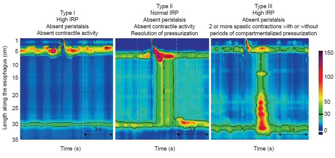
Figure 3. Source: Torresan F., Ioannou A., Azzaroli F., Bazzoli F. Treatment of achalasia in the era of high-resolution manometry. Annals of Gastroenterology.2015;28(1):1–8.
Manometric techniques available in clinical practice include conventional catheters with pressure sensors spaced 3–5 cm apart (solid-state or water-perfused systems), as well as high-resolution manometry (HRM) systems with sensors positioned at 1-cm intervals. All current manometric systems can be used to assess lower esophageal sphincter (LES) relaxation.
Esophageal pressure topography obtained via HRM has enabled classification of achalasia into three subtypes according to the Chicago Classification:
- Type I — achalasia with complete absence of peristalsis;
- Type II — achalasia with panesophageal pressurization;
- Type III — spastic achalasia.
Several retrospective cohort studies have demonstrated that type II achalasia is associated with the best treatment outcomes, whereas type I shows less favorable results and type III is often the most refractory to therapy.
Although these subtypes can be identified using conventional manometry, their differentiation is more accurate and practical with HRM.
Barium esophagography demonstrates characteristic features of achalasia, including esophageal dilation, narrowing at the gastroesophageal junction (“bird’s beak” appearance), absent peristalsis, and impaired contrast emptying.
It is particularly useful in cases where manometric findings are inconclusive. In addition to confirming the diagnosis, barium studies allow assessment of advanced morphological changes such as esophageal tortuosity, angulation, and megaesophagus, which are important for treatment planning.
An additional advantage of barium studies is the objective evaluation of esophageal emptying after treatment, as symptomatic improvement does not always correlate with functional recovery.
The primary role of esophagogastroduodenoscopy (EGD) is to exclude mechanical obstruction or pseudoachalasia, which may mimic achalasia both clinically and manometrically.
Clinical features such as dysphagia combined with advanced age, significant weight loss, and short symptom duration may raise suspicion for malignancy; however, these findings are not specific. Therefore, all patients with suspected achalasia should undergo EGD to exclude cancer.
Endoscopy may also aid in identifying achalasia in patients initially misdiagnosed with gastroesophageal reflux disease (GERD). Typical findings include esophageal dilation with retained food or saliva and narrowing at the gastroesophageal junction. Endoscopic findings may range from a normal-appearing esophagus to a markedly dilated, tortuous (“sigmoid-shaped”) esophagus with food stasis. Consequently, EGD lacks sensitivity in early-stage disease.
The esophageal mucosa is often normal; however, in advanced cases, inflammatory changes or ulcerations may be present secondary to stasis, medication-induced esophagitis, or fungal infection.
In summary, esophageal manometry, EGD, and barium esophagography play complementary roles in the diagnosis of achalasia. EGD is essential for excluding pseudoachalasia, whereas manometry and barium studies are primarily used to confirm the diagnosis.
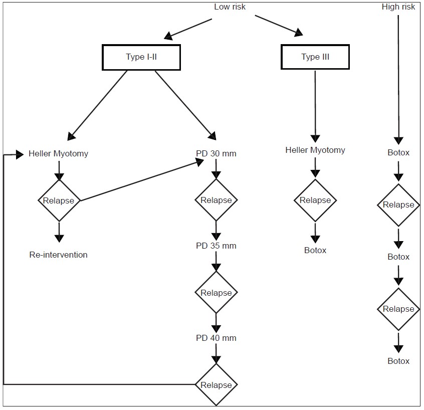
Figure 4.
Source: Torresan F., Ioannou A., Azzaroli F., Bazzoli F. Treatment of achalasia in the era of high-resolution manometry. Annals of Gastroenterology. 2015;28(1):1–8.
TREATMENT
Achalasia is a chronic, incurable disorder. Current treatment strategies are aimed at reducing lower esophageal sphincter (LES) hypertonicity through pharmacological, endoscopic, or surgical interventions. None of the available therapies restore normal esophageal peristalsis, and despite treatment, LES hypertonicity tends to recur over time, necessitating repeated interventions. The primary goals of treatment are symptom relief, improvement of esophageal emptying, and prevention of progressive esophageal dilation. To achieve these goals, the spectrum of available therapeutic options should be individualized according to patient characteristics and clinical indications, following established management algorithms (Fig. 4).
Pharmacologic Therapy
Oral pharmacologic therapy represents the least effective treatment option for achalasia. The two most commonly used drug classes are calcium channel blockers and long- acting nitrates. These agents temporarily reduce lower esophageal sphincter (LES) pressure by inducing smooth muscle relaxation, thereby facilitating esophageal emptying.
Phosphodiesterase-5 inhibitors, such as sildenafil, have also been shown to reduce LES tone in patients with achalasia. Less commonly used agents include anticholinergics, β- adrenergic agonists (e.g., terbutaline), and theophylline.
Calcium channel blockers reduce LES pressure by approximately 13–49%, with symptomatic improvement reported in 0–75% of patients. Nifedipine is the most commonly used agent, achieving peak effect within 20–45 minutes and a duration of action of 30–120 minutes. For optimal efficacy, it is administered sublingually at a dose of 10–30 mg, 30–45 minutes prior to meals.
Isosorbide dinitrate also effectively reduces LES pressure by 30–65%, resulting in symptom improvement in 53–87% of patients. It has a faster onset of action (3–27 minutes) but a shorter duration (30–90 minutes), and is typically administered sublingually at a dose of 5 mg, 10–15 minutes before meals.
A single comparative study of sublingual nifedipine and isosorbide dinitrate demonstrated a non-significant trend toward greater LES pressure reduction with isosorbide dinitrate (65% vs. 49%).
The main limitations of pharmacologic therapy include short-lived clinical response and frequent adverse effects such as headache, hypotension, and peripheral edema. Additionally, these agents do not provide complete symptom relief.
Therefore, pharmacologic therapy is generally reserved for patients who are not candidates for more definitive interventions (pneumatic dilation or surgical myotomy), or in those who have failed botulinum toxin therapy.
Endoscopic Pharmacologic
Therapy Botulinum toxin (Botox) is a potent inhibitor of presynaptic acetylcholine release from nerve terminals and has been shown to be effective in the treatment of achalasia. Its mechanism of action involves cleavage of the SNAP-25 protein, which inhibits acetylcholine exocytosis into the synaptic cleft, resulting in temporary chemical denervation of muscle fibers. This leads to inhibition of cholinergic stimulation of the lower esophageal sphincter (LES), which, in achalasia, is not counterbalanced by inhibitory neural input.
However, botulinum toxin does not affect the myogenic component of basal LES tone, thereby limiting the duration of its therapeutic effect. Clinical efficacy is associated with approximately a 50% reduction in LES pressure, which may be sufficient to allow esophageal emptying when intraluminal pressure exceeds LES pressure.
The main advantages of this approach are its technical simplicity (comparable to standard endoscopy) and a low rate of serious complications. Short-term efficacy is high (>75% at 1 month), but declines to 35–41% at 12 months.
A significant proportion of patients experience symptom recurrence, requiring repeat injections at intervals of 6–24 months (approximately 50% of cases). A more durable response is often observed in elderly patients and may also be associated with type II achalasia.
Serious adverse events are rare. The most common complication (16–25%) is chest pain. Rare complications include mediastinitis and allergic reactions related to protein components.
Repeated injections may induce inflammatory changes extending into the muscular layer, potentially complicating subsequent surgical myotomy. There is also evidence suggesting increased technical difficulty of surgery following botulinum toxin injections.
Given these limitations, botulinum toxin is primarily reserved for patients with high surgical risk in whom pneumatic dilation or surgical myotomy is contraindicated.
Additionally, botulinum toxin may be used as adjunctive therapy in patients with residual spastic contractions above the myotomy site or at the level of the LES.
Pneumatic Dilation
Pneumatic dilation (PD) is the most effective non-surgical treatment option for achalasia. All patients considered for PD should also be candidates for surgical intervention in case of complications, particularly esophageal perforation.
The mechanism of action involves pneumatic expansion of the lower esophageal sphincter, resulting in disruption of the circular muscle fibers, reduced sphincter pressure, and improved esophageal emptying.
The procedure is typically performed under general anesthesia and traditionally under fluoroscopic guidance. However, direct endoscopic guidance for balloon positioning has also been shown to be effective. Key requirements for safe performance include: operator expertise and availability of immediate surgical backup.
Following dilation, all patients should undergo contrast radiography using a water- soluble agent (e.g., Gastrografin), followed by barium esophagography if no perforation is detected. PD can be performed in an outpatient setting, and patients may be discharged after the procedure if clinically stable. Patients must be clearly instructed to seek immediate medical attention if they develop severe chest pain, with or without fever, after discharge.
Surgical Myotomy
Surgical myotomy (Heller myotomy) is considered the gold standard for the treatment of achalasia. It involves division of the circular muscle fibers of the lower esophageal sphincter while preserving the mucosal layer.
Historically, the procedure was performed via a transthoracic approach, achieving good to excellent outcomes in 60–94% of cases with durability ranging from 1 to 36 years. Over time, open approaches were largely replaced by minimally invasive techniques. While thoracoscopic myotomy has been used successfully, laparoscopic myotomy has become the preferred approach due to reduced invasiveness and faster recovery.
Reported outcomes across approaches: оpen transthoracic myotomy — ~83% (64–97%) symptom improvement; оpen abdominal myotomy — ~85% (48–100%); thoracoscopic myotomy — ~78% (31–94%); Laparoscopic myotomy — ~89% (77–100%). However, long-term efficacy declines over time, with symptom relief decreasing from 89% at 6 months to 57% at 6 years.
Gastroesophageal reflux disease (GERD) is a common complication following surgical myotomy and remains a subject of ongoing debate regarding the necessity of prophylactic antireflux procedures. This issue is particularly relevant given the potential increase in postoperative dysphagia associated with fundoplication.
The average incidence of GERD after myotomy without fundoplication is: transthoracic — 29%; open abdominal — 28%; thoracoscopic — 28%; laparoscopic — 31%. The addition of fundoplication significantly reduces GERD incidence: transthoracic — 14%; open abdominal — 8%; laparoscopic — 9%. Data regarding fundoplication following thoracoscopic myotomy remain limited.
The benefit of adding fundoplication was clearly demonstrated in a randomized double-blind trial comparing myotomy with and without an antireflux procedure. Pathological acid exposure on pH monitoring was detected in 47% of patients without fundoplication and in only 9% of those who underwent Dor fundoplication. Subsequent cost- effectiveness analysis based on this trial showed that myotomy combined with Dor fundoplication is more cost-effective than myotomy alone due to reduced long-term GERD- related treatment costs. Current guidelines from the Society of American Gastrointestinal and Endoscopic Surgeons (SAGES) recommend routine addition of fundoplication in patients undergoing myotomy to prevent reflux.
However, the optimal type of fundoplication remains uncertain. The most commonly used techniques are: anterior (Dor) fundoplication and posterior (Toupet) fundoplication. There is no conclusive evidence demonstrating superiority of one technique over the other. Despite fundoplication, some patients may continue to experience reflux symptoms or abnormal pH findings, in which case proton pump inhibitor therapy is indicated. Peroral
Endoscopic Myotomy (POEM)
Peroral endoscopic myotomy (POEM) is a modern minimally invasive technique for the treatment of achalasia based on the principles of natural orifice transluminal endoscopic surgery (NOTES).
The procedure involves creating an entry into the submucosal space by performing a longitudinal mucosal incision (approximately 2 cm), followed by the formation of a submucosal tunnel extending to the level of the lower esophageal sphincter (LES). Through this tunnel, selective myotomy of the circular muscle fibers is performed, typically extending more than 7 cm along the esophagus and approximately 2 cm into the proximal stomach.
Initial studies by Inoue et al. demonstrated a 100% technical and clinical success rate in a small cohort, with significant reduction in LES pressure. Subsequent studies have confirmed high efficacy rates (85–100%), including in patients with prior failed pneumatic dilation.
However, most available data are limited by short follow-up periods (typically up to 6 months), which restricts conclusions regarding long-term outcomes.
A major limitation of POEM is the absence of an antireflux procedure, resulting in a relatively high incidence of gastroesophageal reflux disease, reported in up to 40–46% of patients.
Esophagectomy
In some patients, “end-stage” achalasia may develop, characterized by megaesophagus or a sigmoid-shaped esophagus, with significant dilation and tortuosity.
In this group of patients, pneumatic dilation may be less effective; however, surgical myotomy may be considered a reasonable initial approach before addressing the need for esophagectomy. Two recent studies have documented symptomatic improvement after myotomy in 92% and 72% of patients with megaesophagus.
Nevertheless, in cases of treatment failure, esophageal resection is often required.
Esophagectomy is associated with higher morbidity and mortality compared to laparoscopic Heller myotomy and should be reserved for patients in whom pneumatic dilation and/or myotomy have been ineffective and who are suitable candidates for surgery.
Data from uncontrolled studies indicate generally good outcomes after esophagectomy, with symptom improvement in more than 80% of patients with end-stage achalasia; reported mortality rates range from 0 to 5.4%.
There is a lack of studies comparing the two main approaches to esophagectomy, namely gastric and colonic interposition. However, a recent comprehensive review on this topic demonstrated that gastric interposition is the preferred method in most patients undergoing esophagectomy.
